3 February, 2025
CW tunable lasers and their use in TERS
Photonics research undertakes considerable efforts to continuously refine nanoimaging techniques driven by the desire to characterize electronic and vibronic properties of new materials with nanometer resolution.
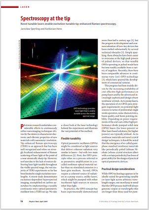
Tip-enhanced Raman spectroscopy (TERS) is an approach that has been well recognized and relies on strongly localized enhancement of Raman scattering of laser light at the point of a near atomically sharp tip. However, not least due to the lack of sources delivering laser light tunable throughout the visible spectral range, the vast majority of TERS experiments so far has been limited to single excitation wavelengths. A recent study demonstrates excitation-dependent hyperspectral imaging, exemplified on carbon nanotubes by implementing a tunable continuous-wave optical parametric oscillator (C-WAVE) into a TERS set-up.
Excitation-Tunable Tip-Enhanced Raman Spectroscopy
The three main components of a TERS set-up a: A laser light source for excitation, an atomic force microscope (AFM) equipped with a carefully selected sharp metallic tip, and a Raman spectrometer recording the inelastically scattered radiation. In essence, the physical principle behind TERS relies on so-called localized surface plasmons that are excited by the laser light in the microscope tip. These plasmons generate a strongly localized electromagnetic field, which not only enhances the incoming and Raman scattered radiation by orders of magnitude, but also ensures a highly localized excitation of the sample under study. Thus, by recording tip-enhanced Raman spectra intensities as a function of the tip position, TERS allows for nanoimaging with a spatial resolution down to below 10 nm.
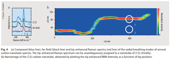
The figure above illustrates the sequence of events and results of a TERS experiment carried out at a single laser excitation wavelength (633 nm) on a film of a CNT mixture. In a first step, a so-called composed Raman spectrum is recorded by placing the microscope tip at a particular (x,y) position in close proximity to the CNT film. The resulting composed Raman spectrum encompasses the RBM peaks of several CNTs (all of them in electronic resonance to the excitation wavelength). In a second step, the microscope tip is retracted, and the so-called far field spectrum is recorded without the tip-enhanced Raman contribution to the signal. By subtracting the far-field spectrum from the composed spectrum, the pure tip-enhanced Raman spectrum is obtained. Eventually, from the pure tip-enhanced Raman spectrum, the tube species underneath the tip position can be unambiguously identified (CNT with (7,5) chirality in the example shown in Figure 4a). For a spatial image of the particular CNT, the outlined procedure is repeated: The tip position is scanned step-wise over the sample surface, and at each point the intensity of the pure tip-enhanced Raman peak is determined. Figure 4b shows the result of such a scan and images the position of a (7,5) CNT in a 2 x 2 μm2 area. As can be clearly perceived, the CNT is around 800 nm long and bent in a step-like shape.
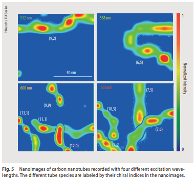
The full beauty of the experimental approach now unfolds when realizing that the imaging capability of the set-up is no longer limited to a subset of CNTs that happen to be in electronic resonance to a particular excitation wavelength. This hyperspectral principle of nanoimaging is at the very heart of the excitation-tunable tip-enhanced Raman spectroscopy (e-TERS) methodology very recently reported by Kusch and co-workers [5]. By using four different excitation wavelengths, they are able to identify and image a total of nine different CNT species within one and the same sample area as show in the images below.
The experimental demonstration of excitation-tunable tip enhanced Raman spectroscopy comes in tandem with the availability of novel tunable laser light sources based on OPO technology. From the general laser technology point-of-view, the performance characteristics of OPOs make them competitive alternatives to conventional lasers and related technologies for the generation of widely tunable cw radiation. From the experimental methodology point-of-view, we expect e-TERS to open new experimental horizons for studying the electronic and vibronic properties of matter on the nanometer scale.
Download the full article for free
The authors gratefully acknowledge enlightening discussions and support by Patryk Kusch from the group of Stephanie Reich at the Free University Berlin.
[5] N. S. Mueller, S. Juergensen, K. Höflich, S. Reich, and P. Kusch, Excitation-Tunable Tip-Enhanced Raman Spectroscopy, J. Phys. Chem C, accepted
More resources
Explore our Publications for practical insights on how our customers are leveraging the power of our lasers in their projects.
Customer publications
Application: Raman spectroscopy
Product line: C-WAVE
Wavelength: Tunable
Optimizing Resonant Raman Scattering in Monolayer
Optimizing the sensitivity of Raman spectroscopy by engineering carrier relaxation dynamics to enhance Resonance Raman scattering intensity
Customer publications
Application: Raman spectroscopy
Product line: Cobolt
Wavelength: 785 nm
Tweaking Bleach’s PH Against Bacterial Spores
Scientists identify the optimal pH for hypochlorite-based sporicidal formulations that can balance solution stability and effectiveness of spore disinfection.
Customer publications
Application: Fluorescence microscopy
Product line: Cobolt
Wavelength: 473 nm, 561 nm
Cobolt Blues and Jive in Uncoventional Protein Secretion
Researchers have identified a specialized compartment called CUPS (Compartment for Unconventional Protein Secretion) that forms independently of the traditional cell pathway system


