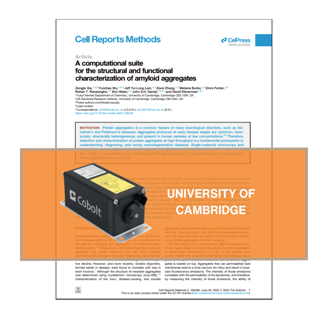
A new tool to automate the analysis of fluorescence microscopy images
A computational suite for the structural and functional characterization of amyloid aggregates
Researchers from the University of Cambridge have unveiled a computational suite designed for the structural and functional characterization of amyloid aggregates. According to experts, amyloid aggregation is a hallmark of several degenerative diseases affecting the brain or peripheral tissues, whose intermediates (oligomers, protofibrils) and final mature fibrils display different toxicity (Protein folding and aggregation into amyloid).
The newly developed tool called Amyloid Characterization Toolkit (ACT) automates the analysis of fluorescence microscopy images, providing precise counting and characterization of amyloid aggregates. For the imaging setup, the scientists employed the used of our Cobolt Jive™ 561nm and our Cobolt 06-MLD 638 nm lasers. With a 90% counting accuracy and rapid processing capabilities, ACT promises to streamline research on amyloid diseases and protein aggregation.
Significance of the study:
This toolkit enhances the ability of researchers to study the formation, structure, and function of amyloid aggregates, providing valuable insights into their role in neurodegenerative diseases. By facilitating more accurate and efficient analysis, ACT can contribute to the development of new therapeutic strategies for conditions like Alzheimer’s disease.
More resources
Looking for more in-depth information? Visit our Knowledge Bank page for detailed articles and insights on our products and technologies.


