News

Multi-line lasers for light sheet microscopy

Over the last decade, the fluorescence-based life science industry has already been transitioning from bulky gas-laser sources into solid-state lasers with a smaller footprint, longer lifetime, and lower maintenance requirements. The development of compact, reliable solid-state lasers was an initial enabling technology for commercialization and expansion to new markets and applications, accompanied by parallel improvements in data storage and advanced camera systems, to name a few. While some applications are able to utilize the advancements in LED and super-continuum white-light sources; the high-resolution, high-speed techniques still rely on the high-brightness and wavelength precision of lasers.

As new techniques are developed for clinical applications, ease-of-use and the ability to commercialize the instrumentation become increasingly important. While maintaining the highest quality and performance, laser manufacturers must deliver reliable, simple, and cost-effective solutions for both commercial systems and laboratory instrumentation or fundamental research.

Why multi-line lasers?

A revolutionary concept made possible thanks to the priority and unique manufacturing method from Cobolt, called HT Cure. A Multi-line laser is a single compact laser head with multiple lasers built inside and collimated into a single permanently aligned output beam, thus giving the appearance of a single laser with multiple wavelengths. The multi-line laser is therefore very attractive as an extremely compact, an easy to use solution for adding more functionality and wavelengths either to a system design or lab set-up. The availability of a compact, easy-to-use, and reliable high-performance multi-line laser will assist with the commercialization of new fluorescence-based-instrumentation and further expand existing technologies into laboratories with a lower barrier of entry, for both the manufacturer and end-user. It is the simplicity and compactness which makes the multi-line laser very attractive for use in light sheet microscopy.

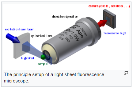
In light sheet microscopy the sample is illuminated by a plane of light instead of a point focus which means that it is exposed to lower doses of light and therefore is less likely to suffer from photothermal damage. For this reason it is considered more of a gentle microscopy method and is thus ideally suited for investigation of living samples.

Light sheet microscopy set upco. Wikipedia

The availability of multi-line lasers enables smaller and more cost-efficient multicolor light sheet microscopy based systems which are easier to manufacture and maintain. This supports the drive to bring more advanced laser-based microscopy techniques into clinical settings for improved medical diagnostics and the further development of new analytical techniques.

To understand the advantages of a multi-line laser watch our flash talk from Focus on Microscopy 2021:

If you’re curious to learn about the impressive work and the potential of light sheet microscopy in clinical settings for improved cancer diagnosis and treatment you’ll enjoy our editorial as seen in BioPhotonics Magazine May/June 2021 issue.

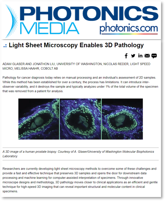
Light Sheet Microscopy Enables 3D pathology

Through innovative microscope designs and methodology, 3D pathology moves closer to clinical applications as an efficient and gentle technique for high-speed 3D imaging that can reveal important structural and molecular content in clinical specimens. Here using light sheet microscopy along with the multi-line laser Cobolt Skyra.

Light sheet enables 3D pathology

2024-10-18T15:44:38+02:00

Multi-line lasers simplify fluorescence microscopy

Editorial: Photonics Views June 2019 edition
Multi-line Lasers simplify fluorescence microscospy

by Melissa Haahr

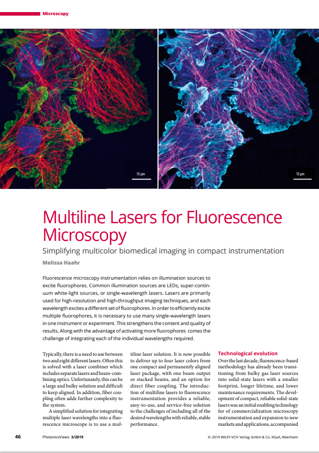
Fluorescence microscopy instrumentation relies on illumination sources to excite fluorophores. Common illumination sources are LEDs, super-continuum white-light sources, or single-wavelength lasers.Lasers are primarily used for high-resolution and high-throughput imaging techniques, and each wavelength excites a different set of fluorophores. In order to efficiently excite multiple fluorophores, it is necessary to use many single-wavelength lasers in one instrument or experiment.  This strengthens the content and quality of results. Along with the advantage of activating more fluorophores comes the challenge of integrating each of the individual wavelengths required.

Technological evolution

Over the last decade, fluorescence-based methodology has already been transitioning from bulky gas laser sources into solid-state lasers with a smaller footprint, longer lifetime, and lower maintenance requirements. The development of compact, reliable solid-state lasers was an initial enabling technology for of commercialization microscopy instrumentation and expansion to new
markets and applications, accompanied by parallel improvements in data storage and advanced camera systems, to name a few. While some applications are able to utilize the advancements in LED and super-continuum white-light sources, the high-resolution, high-speed techniques still rely on the high brightness and wavelength precision of lasers.

Currently, many researchers and manufacturers align and integrate individual laser sources for each wavelength on the optical bench or in instrument. These laser combiners and laser light engines have simplified some of these assemblies substantially. However, they do not eliminate the need for alignment (and re-alignment) over time. A permanently aligned multi-line laser offers a robust and alignment free option.

Multi-line laser technology

The Cobolt Skyra multi-line laser is unique in its design and manufacturing. It is built using patent-pending alignment techniques and utilizing Cobolt’s proprietary HTCure technology. The HTCure technology is based on careful thermo-mechanical matching and high-temperature fixation of miniaturized optics. The lasers are built on a single, temperature-controlled platform for stable operation and protection from thermomechanical misalignment.

Skyra fluorescence microscopy

The Skyra can include up to four wavelengths, within the range of 405 nm to 660 nm with beam position overlap <50 μm at the exit and pointing stability <10 μrad/°C over a temperature range of 20 °C to 50 °C.

Read the full article on PHOTONICS Views web site.

lasers fluorescence microscopy

2024-10-03T09:13:41+02:00

Multi-line lasers for microscopy

Why choose a multi-line laser for microscopy instead of individual lasers?

Conventional fluorescence-based bio-instrumentation equipment typically uses multiple individual lasers combined through optical elements into one beam or an optical fiber. The systems can become bulky, costly to manufacture, and challenging to keep aligned. An extremely compact, permanently aligned, and service-free multi-line laser device can reduce the size and cost of these systems for fluorescence-based research. Removing the complexity of integrating individual lasers and replacing with a single multi-line laser solution allows the techniques to be more cost-efficient, user-friendly, and accessible for all levels of researchers.

Fluorescence microscopy instrumentation relies on illumination sources to excite fluorophores. Common illumination sources are LEDs, super-continuum white-light sources, or single-wavelength lasers. Lasers are primarily used for high-resolution and high-throughput imaging techniques and each wavelength excites a different set of fluorophores. In order to efficiently excite multiple fluorophores, it is necessary to use many single-wavelength lasers in one instrument or experiment.

Along with the advantage of activating more fluorophores, comes the challenge of integrating each of the individual wavelengths required. Typically, there is a need to use between two and eight different lasers. Often this is solved with a laser-combiner, which includes separate lasers and beam-combining optics. Unfortunately, laser combiners can be a large and bulky solution, and difficult to keep aligned. In addition, fiber coupling often adds further complexity to the optical system.

A simplified solution for integrating multiple laser wavelengths into a fluorescence microscope is to use a multi-line laser solution. It is now possible to deliver up to four laser colors from one compact and permanently aligned laser package, with one beam output or stacked beams, and an option for direct fiber coupling [1]. The introduction of multi-line lasers to fluorescence instrumentation provides a reliable, easy-to-use, and service-free solution to the challenges of including all of the desired wavelengths with reliable, stable performance.

Cobolt Skyra™ – Multi-line Laser

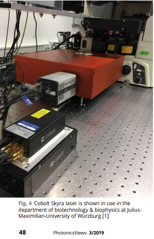
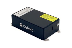
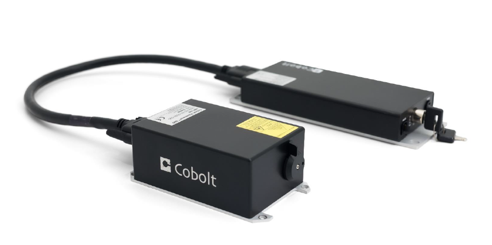
The compact Cobolt Skyra multi-line laser

In addition to the simplification of laser integration, a multi-line laser also brings the advantage of customization and unique control capabilities. Across different techniques, laboratories, or even individual experiments, there are various requirements on the colors, power output, modulation, and beam configuration; all of which can now be accommodated with one ultra-stable solution.

[1] SPIE Paper 10884-35 Fluorescence microscopy simplified using novel multi-line lasers – MHaahr

2024-10-03T09:13:37+02:00

Transient State Imaging microscopy (TRAST) for cancer diagnosis

Transient State Imaging microscopy (TRAST) for cancer diagnosis

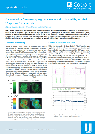
Application note: Transient State Imaging or TRAST microscopy for measuring oxygen concentration in cancerous cells.

In the field of biology it is a general consensus that cancerous cells often use other metabolic pathways, than corresponding healthy cells, and thereby consume less oxygen. If it is possible to measure the oxygen levels of cells by fluorescence microscopy this could be exploited as a future tool in clinical cancer diagnosis. However, measuring oxygen concentration of live cells is not totally straight forward. Traditional fluorophores have too short excited state lifetimes (nanoseconds) to be significantly influenced by molecular oxygen collisions, typically taking place in the microsecond time range.

A new technique called Transient State Imaging (TRAST) is set to change the way oxygen concentration of cells is measured, by taking advantage of transitions to and from the dark lowest triplet state (T1) of fluorophores (1). T1 is a photo-induced, long-lived non-fluorescent state, found in essentially all fluorophore molecules. Combining fluorescence microsocpy with a modulated laser source, and systematically varying the modulation characteristcs, it is possible to extract kinetic information about the T1 state.

Read the full application note for all the details:

TRAST imaging microscopy

Share this article!

2024-10-03T09:13:30+02:00

640 nm for STORM microscopy

High power 640 nm lasers make STORM microscopy easier

HÜBNER Photonics introduces the Cobolt Rogue™ 640 nm laser, especially for super resolution STORM microscopy. The Cobolt Rogue™ Series lasers are continuous-wave diode pumped lasers (DPL) and are multi-mode, high power complements to our Cobolt 05-01 Series of single frequency lasers. The Cobolt Rogue™ 640 nm is multi-longitudinal mode in a perfect TEM00 beam with 1 W output power, ideally suited for Single Molecule Localization Microscopy (SMLM) including super resolution STORM microscopy.

In SMLM, high irradiance (several kW/cm²) is crucial to reach a single molecule regime and hence assuring quantitative imaging at the nanoscale. It becomes even more critical while imaging simultaneously 3 fluorophores at the density needed for SMLM over large fields of view (FOV). Furthermore, as higher irradiance increases the blinking rate, increased irradiance allows for faster imaging at the nanoscale.

The need for high irradiance to excite multiple fluorophores with nanoscale resolution over large FOVs and at high speeds translates into the need for high power laser sources. In this regard, the availability of new compact and powerful laser sources in the red spectral range is facilitating the development of simplified multicolor SMLM imaging technology with high throughput capability. The new 1W CW laser at 640 nm, the Cobolt Rogue, is suitable for simultaneous excitation of red dyes, as AF647 and CF680. The Cobolt Rogue is a diode-pumped laser emitting directly at 640 nm as the fundamental wavelength and emitting a diffraction limited, single-transversal mode TEM00 beam, which enables efficient coupling of the emission into single-mode polarization maintain fibers with consequent easy and efficient coupling of the light into the microscope.

All Cobolt lasers are manufactured using proprietary HTCure™ technology and the resulting compact hermetically sealed package which provides a very high level of immunity to varying environmental conditions along with exceptional reliability.

With demonstrated lifetime capability and several thousand units installed in the field, Cobolt lasers have proven to deliver unmatched reliability and performance both in laboratory and industrial environments, and are offered with market leading warranty terms.

Read more  about the Cobolt Rogue HERE

2024-10-03T09:13:24+02:00
Go to Top