Deprecated: Array and string offset access syntax with curly braces is deprecated in /var/www/vhosts/hubner-photonics.enestedt-playground.se/httpdocs/wp-content/plugins/woocommerce-wootabs/public/class-wootabs_wp_editor.php on line 181

Deprecated: Array and string offset access syntax with curly braces is deprecated in /var/www/vhosts/hubner-photonics.enestedt-playground.se/httpdocs/wp-content/plugins/woocommerce-wootabs/public/class-wootabs_wp_editor.php on line 181

Deprecated: Array and string offset access syntax with curly braces is deprecated in /var/www/vhosts/hubner-photonics.enestedt-playground.se/httpdocs/wp-content/plugins/woocommerce-wootabs/public/class-wootabs_wp_editor.php on line 181

Deprecated: Array and string offset access syntax with curly braces is deprecated in /var/www/vhosts/hubner-photonics.enestedt-playground.se/httpdocs/wp-content/plugins/woocommerce-wootabs/public/class-wootabs_wp_editor.php on line 181
Customer publications Archives - Page 2 of 2 - HÜBNER Photonics

News


Notice: Undefined offset: 0 in /var/www/vhosts/hubner-photonics.enestedt-playground.se/httpdocs/wp-content/themes/Avada-Child-Theme/templates/blog-layout.php on line 157

Notice: Undefined offset: 0 in /var/www/vhosts/hubner-photonics.enestedt-playground.se/httpdocs/wp-content/themes/Avada-Child-Theme/templates/blog-layout.php on line 158

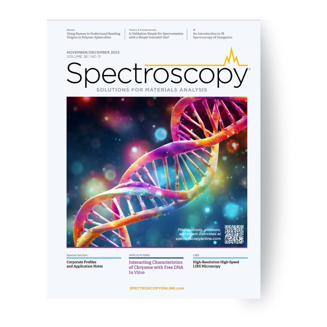
New editorial: High-Resolution High-Speed LIBS Microscopy

We are excited to share our latest featured article in Spectroscopy co-authored by our HÜBNER Photonics product manager Elena Vasileva.

LIBS (Laser-Induced Breakdown Spectroscopy) imaging is revolutionizing our ability to analyze elemental and mineralogical distribution within samples. Traditionally, we’ve relied on excitation sources like lasers with a repetition rate of 10-100 Hz for LIBS analysis. But what if we could enhance our capabilities for high-resolution imaging without enduring lengthy acquisition times?

The research shows how the development of a µ-LIBS imaging microscope can provide an astonishing 10 µm resolution in under 20 minutes per cm². For the first time, we can capture high-resolution images, revealing intricate elemental distribution within the analyzed samples.

The world of LIBS imaging is evolving, and we’re at the forefront of this exciting journey! This opens up endless possibilities across various research fields, from biomedical and geological material analysis to industrial applications like mining!

Read more in the magazine on page 34!

2024-09-23T15:42:22+02:00

Revolutionizing Live-Cell Microscopy: Advancements in Label-Free Imaging Techniques

In the realm of live-cell microscopy, fluorescence techniques have long dominated, yet challenges persist due to bleaching and motion blur caused by extended integration times. However, a breakthrough has emerged with Rotating Coherent Scattering (ROCS) microscopy, enabling high-contrast, label-free imaging of live cells with unprecedented speed and resolution.

Researchers at Freiburg University, Germany, have harnessed the power of our Cobolt 06-01 and Cobolt 05-01 laser series to study cell samples in minute details. ROCS microscopy capitalizes on intensity speckle patterns from all azimuthal illumination directions, aggregating thousands of acquisitions within a mere 10 milliseconds at a remarkable resolution of 160 nm and a rapid capture rate of 100 Hz.

Through sophisticated analysis methods, this research not only reveals how motion blur obscures cellular structures but also elucidates how slow structural motions can mask critical fast motions, offering profound insights into the dynamic processes within living cells.

The study heralds a new era in live-cell microscopy, promising unparalleled discoveries in cellular dynamics.

2024-09-23T15:42:22+02:00

Exploring Readout Contrasts: Tuning Optical and Electrical Measurements of NV Centers in Diamond

In a recent investigation, researchers at the Munich Center for Quantum Science and Technology, in collaboration with the Technical University of Munich in Germany use our C-WAVE Tunable laser to delve into the nuanced contrasts observed in the electrical and optical readout of NV centers in diamond, shedding light on their dependence on optical excitation wavelength and various excitation schemes.

The study shows that while optically detected magnetic resonance (ODMR) showcases efficient performance within the 480 to 580 nm range, electrically detected magnetic resonance (EDMR) exhibits a pronounced reliance on excitation dynamics. Remarkably, the study uncovers that the most substantial electrically detected contrast, reaching -23%, is attained by resonantly exciting the zero-phonon line of the neutral charge state of NV at 575 nm.

These findings offer crucial insights into the intricacies of NV center behavior, paving the way for refined techniques in both optical and electrical readout methodologies.

2024-09-23T15:42:23+02:00

Harnessing Ultrafast Fluctuations for Enhanced Nonlinear Imaging

Researchers in Japan have uncovered a pivotal aspect of optical parametric generators: the phenomenon of bunching caused by ultrafast intensity fluctuations. This mechanism, previously recognized for enhancing nonlinear interactions between light and matter, has now been investigated in the context of sufficiently intense light pulses crucial for biological nonlinear imaging.

By harnessing the power of our Cobolt Tor™ Series, the scientists showcase the enhanced two-photon excited fluorescence enabled by ultrafast fluctuations within intense pulses.

Armed with this calibrated optical parametric generator, the study achieved a breakthrough in two-photon imaging of green fluorescent protein within brain tissue, all within a mere timescale of seconds. These experimental triumphs underscore the immense potential of intense pulses and the bunching effect induced by ultrafast fluctuations, offering a promising avenue for advancing nonlinear imaging in the realms of biology and medicine.

This discovery promises to revolutionize our understanding and application of nonlinear imaging techniques, paving the way for transformative breakthroughs in biomedical research and clinical diagnostics.

2024-09-23T15:42:23+02:00
Go to Top