Deprecated: Array and string offset access syntax with curly braces is deprecated in /var/www/vhosts/hubner-photonics.enestedt-playground.se/httpdocs/wp-content/plugins/woocommerce-wootabs/public/class-wootabs_wp_editor.php on line 181

Deprecated: Array and string offset access syntax with curly braces is deprecated in /var/www/vhosts/hubner-photonics.enestedt-playground.se/httpdocs/wp-content/plugins/woocommerce-wootabs/public/class-wootabs_wp_editor.php on line 181

Deprecated: Array and string offset access syntax with curly braces is deprecated in /var/www/vhosts/hubner-photonics.enestedt-playground.se/httpdocs/wp-content/plugins/woocommerce-wootabs/public/class-wootabs_wp_editor.php on line 181

Deprecated: Array and string offset access syntax with curly braces is deprecated in /var/www/vhosts/hubner-photonics.enestedt-playground.se/httpdocs/wp-content/plugins/woocommerce-wootabs/public/class-wootabs_wp_editor.php on line 181
Cobolt Archives - HÜBNER Photonics

News


Notice: Undefined offset: 0 in /var/www/vhosts/hubner-photonics.enestedt-playground.se/httpdocs/wp-content/themes/Avada-Child-Theme/templates/blog-layout.php on line 157

Notice: Undefined offset: 0 in /var/www/vhosts/hubner-photonics.enestedt-playground.se/httpdocs/wp-content/themes/Avada-Child-Theme/templates/blog-layout.php on line 158

New Discovery in Quantum Technology: Shining Light on Organic Molecules

Reversible spin-optical interface in luminescent organic radicals

Scientists have achieved a significant advancement in quantum information science by developing organic molecules that exhibit both efficient luminescence and near-unity generation yield of excited states with spin multiplicity S > 1. This discovery could lead to better ways of sensing and computing using the principles of quantum physics.

Historically, carbon-based quantum candidates have been non-luminescent, hindering optical readout via emission. However, the new organic molecules designed by researchers overcome this limitation.

The secret lies in a special design that allows these molecules to switch between different energy states quickly and efficiently. By linking certain chemical groups together, the researchers created a system where energy moves rapidly within the molecule, leading to a bright glow and stable high-spin states, which are crucial for quantum technology. The method supports high efficiency of initialization, spin manipulations, and light-based readout at room temperature. The experiment was conducted with the help of the Cobolt Samba CW 532 nm DPSSL laser.

The team behind the research is a group of scientists hailing from 9 different Universities and research institurions. These include the Universities of Cambridge, Oxford, Manchester and Swansea in the United Kingdom, Wüzerbug in Germany, Jilin in China, the University of Mons and the University of Namur in Belgium, and the Donostia International Physics Center Foundation in Spain. This was a noteworthy international collaborative effort which results were published in the international journal of Nature.

The integration of luminescence and high-spin states in organic molecules paves the way for new platforms in emerging quantum technologies, marking a notable stride towards advanced quantum sensing and computation.

2024-12-09T12:09:14+01:00

Nanographene Research Unveils Highly Soluble Quantum Dots

Interplay of structure and photophysics of individualized rod-shaped graphene quantum dots with up to 132 sp² carbon atoms


In a significant stride towards advancing nanotechnology applications, researchers from the Université Paris-Saclay, Chemical and Biological Systems Simulation Lab and the Laboratory for Chemistry of Novel Materials have unveiled a new family of nanographene materials with the help of the Cobolt Mambo 594 nm laser.


These materials, known as 0D graphene quantum dots, have emerged as promising candidates for single quantum emitters due to their exceptional properties and can potentially revolutionize optics, electronics, and biophotonics.


A key challenge in harnessing the full potential of graphene quantum dots lies in ensuring their high purity. Researchers have identified solubility as a critical factor in achieving efficient purification of these materials. Addressing this hurdle, the team of scientists has successfully synthesized a series of rod-shaped graphene quantum dots that exhibit remarkably high solubility and ease of processing.


The breakthrough, detailed in Nature Communications, highlights fluorescence quantum yields reaching up to an impressive 94%, particularly notable for their red emission capabilities. The enhanced solubility of these quantum dots is attributed to their meticulously designed structure, enabling a precise characterization of their photophysical properties both in solution and at the single molecule level.


Crucially, these photophysical properties were accurately predicted through advanced quantum-chemical calculations, underscoring the scientific rigor behind the findings. This development not only paves the way for further exploration in quantum dot applications but also underscores the potential of nanographene materials in shaping future technologies.


2024-12-20T10:53:44+01:00

New Breakthrough in Cellular Imaging with the Cobolt 06-MLD Laser

Super-sectioning with multi-sheet reversible saturable optical fluorescence transitions (RESOLFT) microscopy

Scientists at the KTH Royal Institute of Technology in Stockholm Sweden, in collaboration with Calico Life Sciences, a biotechnology company founded by Google’s parent company Alphabet Inc., have made significant strides in cellular imaging. The work, published in the prestigious academic journal Nature Methods in May 2024 highlights the development of a new method called multi-sheet RESOLFT (reversible saturable optical fluorescence transitions) microscopy, which was achieved with the help of a specialized Cobolt 06-MLD laser. This innovation promises to revolutionize the way we observe and understand subcellular structures in living cells.

The technique improves upon traditional light-sheet fluorescence microscopy (LSFM) by enabling the imaging of fine subcellular details with minimal photodamage and higher speed. By using reversibly switchable fluorescent proteins (RSFPs) and a periodic light pattern, the group was able to generate multiple ultra-thin emission sheets. This method allowed for rapid volumetric imaging, capturing high-resolution images of subcellular structures in living cells.

The Cobolt laser’s precision was instrumental in producing these thin light sheets, crucial for the super-resolution imaging that the study achieved. The laser’s stability and ability to maintain consistent illumination were key factors that enabled the researchers to observe cellular processes such as cell division and actin motion in real-time.

A Step Forward in Imaging Technology

This advancement is not just a technical marvel for academic laboratories but holds significant implications for medical and biological research. By enabling detailed, rapid, and minimally invasive imaging of living cells, the multi-sheet RESOLFT technique can enhance our understanding of cellular processes such as cell division, cytoskeleton dynamics, and virus particle movements. This could lead to better diagnostic tools, improved treatments for diseases, and a deeper understanding of fundamental biological processes.

The team’s success demonstrates the potential of integrating advanced laser technology with innovative imaging techniques. With the Cobolt 06-MLD laser, scientists can now achieve volumetric imaging at unprecedented resolutions, opening new frontiers in the study of life at the microscopic level.

2024-09-23T15:56:08+02:00

Scientists unveil a new tool to automate the analysis of fluorescence microscopy images

A computational suite for the structural and functional characterization of amyloid aggregates

Researchers from the University of Cambridge have unveiled a computational suite designed for the structural and functional characterization of amyloid aggregates. According to experts, amyloid aggregation is a hallmark of several degenerative diseases affecting the brain or peripheral tissues, whose intermediates (oligomers, protofibrils) and final mature fibrils display different toxicity (Protein folding and aggregation into amyloid).

The newly developed tool called Amyloid Characterization Toolkit (ACT) automates the analysis of fluorescence microscopy images, providing precise counting and characterization of amyloid aggregates. For the imaging setup, the scientists employed the used of our Cobolt Jive™ 561nm and our Cobolt 06-MLD 638 nm lasers. With a 90% counting accuracy and rapid processing capabilities, ACT promises to streamline research on amyloid diseases and protein aggregation.

Significance of the study:

This toolkit enhances the ability of researchers to study the formation, structure, and function of amyloid aggregates, providing valuable insights into their role in neurodegenerative diseases. By facilitating more accurate and efficient analysis, ACT can contribute to the development of new therapeutic strategies for conditions like Alzheimer’s disease.

2024-09-23T15:54:00+02:00

Cambridge University’s new findings on Alzheimer’s disease

Super-resolution Imaging Unveils the Self-replication of Tau Aggregates Upon Seeding

Dementia is one of the major causes of disability among older people affecting 50 million people worldwide. Alzheimer’s disease (AD) is the most common form of dementia, characterized by extensive neuronal loss and severe brain shrinkage. As experts have noted, this pathology is driven by the accumulation of extracellular amyloid-beta (Ab) plaques and intracellular hyperphosphorylated tau aggregates. As it turns out, in Alzheimer’s disease, an abnormal form of tau accumulates and begins sticking together in thread-like structures called neurofibrillary tangles. These tangles are not effectively disposed of through the cell’s usual ways of removing “trash.” As a result of this build-up the microtubule superhighway becomes damaged, thus disrupting the normal functions of neurons (Tau protein in Alzheimer’s disease).

Researchers from the University of Cambridge have made significant strides in understanding Alzheimer’s disease by using super-resolution microscopy to observe tau protein aggregates within cells with the help of the Cobolt 06-MLD. The group used the following wavelengths: 405, 488, 561 and 638 nm.

For the first time, scientists are able to visualise how tau aggregates form and replicate. Their study highlights that even without external seeding, cells can spontaneously form small tau aggregates.

Significance of the study:

The research is significant as it clarifies how tau aggregates replicate themselves, which helps advance our knowledge on the mechanisms behind neurodegenerative disease such as Alzheimer’s. Researchers will be able to harness this information for early detection and diagnosis to empower earlier treatment and better patient outcomes for the future.

2024-09-23T15:51:32+02:00

Researchers uncover new insights on bacterial proteins’ transport system

Conformational coupling of the sialic acid TRAP transporter HiSiaQM with its substrate binding protein HiSiaP

Researchers from the University Hospital of Bonn and the University of Bonn in collaboration with the University of York, have uncovered how bacterial proteins work together to transport sialic acid, a crucial nutrient, across cell membranes. The study, published in Nature Communications, focuses on the TRAP (Tripartite ATP-independent Periplasmic)  transporter system, specifically examining the proteins HiSiaQM and HiSiaP. 

In their finding they observed that the TRAP transporter uses a substrate-binding protein (SBP) to capture sialic acid, a sugar molecule. The study identified how certain mutations could trap HiSiaP in a closed state when it binds sialic acid, providing insight into the protein’s shape changes during the transport process.

A notable technique used in the study was disulfide engineering, an important biotechnological tool to study protein dynamics, to “lock” the transporter and SBP in specific states. To visualize the proteins, the scientist used a custom-built, single molecule sensitive, inverted microscope capable of total internal reflection fluorescence (TIRF) microscopy with illumination source provided by our Cobolt 06-MLD 488 nm laser.

Significance of the study:

This research provides detailed insights into how bacterial transport systems work, which is crucial for developing strategies to combat bacterial infections. Understanding how this mechanism work opens up potential avenues for developing new antibiotics that can disrupt this transport system. By inhibiting the TRAP transporter, it might be possible to prevent pathogenic bacteria from acquiring essential nutrients, thereby enhancing the effectiveness of the immune response against these bacteria.

REFERENCES:  ScienceDaily; ​​ Phys.org; ​​ImmunoSensation² ; Disulfide

2024-09-23T15:53:04+02:00

New study: CZTS Nanocrystals Boost Solar Panel Efficiency

A new study published in the journal of Nanomaterials explores the potential of a material known as Cu2ZnSnS4 (CZTS) for use in solar cells and as a potential thermoelectric material. Researchers at Chemnitz University of Technology in Germany, in collaboration with V.E. Lashkaryov Institute of Semiconductor Physics NAS of Ukrain and Taras Shevchenko National University of Kyiv, found that when CZTS is transformed into tiny particles (nanocrystals) and combined with a common polymer called PEDOT:PSS, shows promising results. However, the study reveals a twist: while the combination enhances the thermoelectric properties, it also affects the structural stability of the polymer. The team used Raman spectroscopy and our Cobolt Fandango 515 nm to uncover these insights. This breakthrough could pave the way for more efficient and durable solar panels, but further research is needed to address the stability issues.


2024-11-04T13:12:07+01:00

Waveguide holography for 3D augmented reality glasses – New research

In a significant stride towards the future of augmented reality (AR) and virtual reality (VR), META Reality Labs, in collaboration with Seoul National University, have unveiled a groundbreaking compact holographic near-eye display concept using our Cobolt Samba 532 nm laser. This prototype addresses critical challenges faced by current technologies, promising immersive and comfortable visual experiences for users.

As the researchers discuss in the journal Nature Communications, the concept aims to overcome obstacles such as achieving a compact form factor, resolving vergence-accommodation conflicts, and attaining high resolution with a large eyebox. Traditionally, these challenges have been stumbling blocks in the quest for creating true 3D holographic augmented reality glasses.

The key to this advancement lies in a meticulous approach to modeling coherent light interactions and propagation through a waveguide combiner. The researchers showcase their ability to control the output wavefront by utilizing a spatial light modulator located at the input coupler side. This method facilitates 3D holographic displays through exit-pupil expanding waveguide combiners, providing a generously sized, software-steerable eyebox.

Moreover, the proposed method brings added benefits, including resolution enhancement capabilities achieved by suppressing phase discontinuities resulting from the pupil replication process. The combination of these features positions the holographic near-eye display concept as a promising candidate for the next generation of computing platforms and its potential to impact the future of AR and VR technologies.

As technology continues to advance, the prospect of true 3D holographic augmented reality glasses draws closer to reality.

2024-09-23T15:54:46+02:00

New Approach Unveiled: Harnessing Dark Spins for Quantum Computing

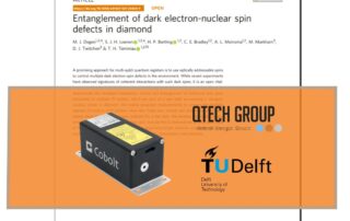
In a groundbreaking development for quantum computing, researchers at Delft University of Technology in the Netherlands, in collaboration with Sweden-based manufacturing company QTECH Group, have unveiled a promising strategy for multi-qubit quantum registers with our Cobolt 06 MLD 515 nm laser.

By utilizing optically addressable spins to manipulate numerous dark electron-spin defects within the environment, significant progress has been made in controlling these elusive quantum entities. Recent experiments have showcased coherent interactions with dark spins, marking a crucial milestone.

These results not only signify a bold step forward in quantum computing, but they also lay the foundation for utilizing dark electron-nuclear spin defects as qubits for diverse applications, including quantum sensing, computation, and networking.

This heralds a new era in quantum technology, offering promising prospects for revolutionizing information processing capabilities.

2024-09-23T15:42:22+02:00

New editorial: High-Resolution High-Speed LIBS Microscopy

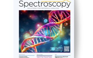
We are excited to share our latest featured article in Spectroscopy co-authored by our HÜBNER Photonics product manager Elena Vasileva.

LIBS (Laser-Induced Breakdown Spectroscopy) imaging is revolutionizing our ability to analyze elemental and mineralogical distribution within samples. Traditionally, we’ve relied on excitation sources like lasers with a repetition rate of 10-100 Hz for LIBS analysis. But what if we could enhance our capabilities for high-resolution imaging without enduring lengthy acquisition times?

The research shows how the development of a µ-LIBS imaging microscope can provide an astonishing 10 µm resolution in under 20 minutes per cm². For the first time, we can capture high-resolution images, revealing intricate elemental distribution within the analyzed samples.

The world of LIBS imaging is evolving, and we’re at the forefront of this exciting journey! This opens up endless possibilities across various research fields, from biomedical and geological material analysis to industrial applications like mining!

Read more in the magazine on page 34!

2024-09-23T15:42:22+02:00
Go to Top